Diaphragmatic Breathing & Neck Pain
Updated on 8/18/21
Diaphragmatic breathing is a pattern of expiration and inspiration in which the diaphragm does most of the ventilatory work. If you observe how babies breathe, you will notice the ease with which their bellies rise and fall. On the flip side, most adults’ breathing patterns are quick and shallow, with very little belly movement and quite a bit of chest elevation.
Everyone from elite athletes to average clients can benefit from learning more about diaphragmatic breathing or reprogramming the way they breathe. More specifically, by teaching them techniques that emphasize diaphragmatic breathing, you will help them meet their exercise goals.
Through years of poor postural habits, myofascial restrictions, stress, trying to “suck in” the abdomen and a host of other factors, we’ve steadily removed our diaphragms from the breathing equation and begun to rely on upper-chest accessory muscles to accomplish this all-important task. Shallow and/or quick breathing (extreme cases are referred to as hyperventilation) is associated with many negative health consequences, including respiratory alkalosis, anxiety, irritable bowel syndrome, decreased pain threshold and allergies. In this discussion, we will focus on the impact such breathing has on core stability and chronic neck and shoulder pain (Chaitow 2004). We will also address assessment methods for identifying poor breathing habits in your clients and review some corrective strategies to help them reprogram their dysfunctional habits.
Diaphragmatic Breathing and Core Stability
Most people consider breathing a relatively rudimentary task, and they may balk at the prospect of spending time retraining it. If the massive amount of evidence linking poor breathing habits to muscle fatigue and decreased performance is not compelling for your clients, perhaps knowing that other movement patterns cannot be normalized until breathing is addressed will get their attention (Lewit 1980).
Diaphragmatic breathing’s effect on movement patterns is chiefly due to the dual role the diaphragm plays in core stabilization and respiration. Traditionally, we have placed the transversus abdominis on a stabilizing pedestal; however, current literature suggests that the diaphragm, transversus abdominis, multifidus and pelvic floor work in unison to create the ideal intra-abdominal pressure for spinal stabilization (Hodges & Gandevia 2000b). These “inner core” muscles fire in an anticipatory manner milliseconds before the prime movers in an effort to stabilize the spine at the segmental level (Hodges & Gandevia 2000b).
Specifically, the diaphragm contributes to spinal stability via a hydraulic effect on the abdominal cavity, thereby increasing intra-abdominal pressure (McGill, Sharratt & Seguin 1995). The problem is that a deconditioned and poorly aligned diaphragm won’t be of much help in providing the pressure and rigidity necessary for the trunk to lift things and move without insult to the spine.
See also: A Strong Diaphragm for a Strong Core
Diaphragmatic Breathing and Posture
As personal trainers, we see postural breakdown coinciding with poor breathing habits all too frequently. Imagine a chest-breathing runner on the treadmill: without adequate participation of the diaphragm, the rib cage tilts posteriorly, the shoulder girdle elevates and the cervical spine hyperextends. This disorganized body, coupled with the perpetual jarring of the foot strike, places the cervical and lumbar spine in an unstable, vulnerable position.
Shallow-breathing runners often experience tension in their neck and commonly suffer from low-back pain. Consider a chest-breathing client performing his or her last set of deadlifts; the body has to choose between stabilizing the low back and maintaining normal respiration (McGill, Sharratt & Seguin 1995). Since respiration trumps every other function in the body, the low back is now at risk. If this were not the case, we’d see people turning blue and passing out in the gym on a regular basis. The simple truth is that we can manage with less-than-ideal breathing habits, but it will be at the expense of spinal stability.
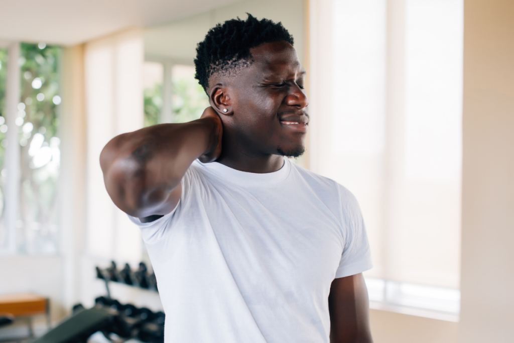
Dysfunctional Breathing and Neck Pain
When the diaphragm’s involvement in breathing decreases, the anterior cervicals (most notably the scalenes) compensate. The scalenes adaptively shorten when they must perform a task they were not designed to do. This becomes problematic because of the potential for the scalenes to entrap the nearby brachial plexus and subclavian artery. Entrapment of these structures is associated with thoracic outlet syndrome, paresthesia, coldness, claudication and lymphedema (Simons, Travell & Simons 1999).
Bilateral tension of the scalenes can also lend itself to forward-head posture, which often results in rounding of the thoracic spine, elevated and protracted shoulders, and scapular winging. Over time, these postural imbalances can result in joint dysfunction at the atlanto-occipital joint, the C4-C5 segment, the cervicothoracic joint, the glenohumeral joint and the T4-T5 segment (Hertiling & Kessler 2006). Forward-head carriage may also promote accelerated aging of intervertebral joints, resulting in degenerative joint disease (White & Sahrmann 1994).
When clients come into the gym with neck and shoulder pain, consider the notion that their problem may actually be stemming from a breathing problem. Although it can be hard to ascertain which came first—dysfunctional breathing or postural distortion—it’s evident that the training protocol needs to address both.
See also: The Many Dimensions of Pain
Assessment
Here are some specific ways to recognize and assess upper-chest breathing:
Posture. Generally speaking, look for a forward head, elevated shoulder girdle, shoulders in a protracted position, an immobile and kyphotic thoracic spine, an anterior tilt of the pelvis and/or a unilateral or bilateral rib flare.
Breathing. The following two breathing assessments are a good place to start an evaluation. Whichever test you choose to administer, observe the length of time for the client to complete a deep breath—a healthy, full inhalation and exhalation should take not less than 10 seconds.
- Fingers and thumbs separation test. Have the client sit in a chair, with you standing behind it. Position your hands so the fingers are facing forward, resting on the superior aspects of the client’s lower ribs, and your thumbs are on the client’s midline posteriorly. Squeeze slightly. Instruct the client to take a deep breath, and note how your fingers and thumbs move. Both the fingers and thumbs should be moving apart from each other, ideally 1.5–2 inches apart (Chaitow et al. 2002).
- The high-low test. Have the client be either supine or in a seated position. Instruct the client to place one hand on the chest, and the other on the belly, and then cue the client to take a few relaxed breaths. During the breaths, take note of positional changes to the client’s hands. Ideally, the hand on the belly should rise before the hand on the chest. The hand on the chest should move slightly forward (not toward the chin). If it moves significantly more than the hand on the abdomen, there is a suggestion of dysfunctional breathing (Simons, Travell & Simons 1999).
A few other telltale signs of dysfunctional breathing are frequent sighs or yawns, repeated throat clearing or air gulping and/or continual mouth breathing.
Even though it may seem less exciting than other protocols, diaphragmatic breathing serves as a basis for functional movement and core stability. Help your clients optimize their diaphragmatic breathing function and you will see them achieve their fitness goals while also reducing some of their nagging aches and pains.